General description
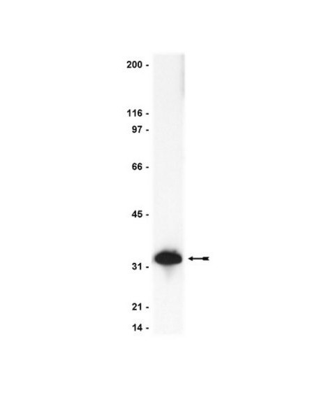
32 kDa
Caspase-3 (UniProt: P42574; also known as EC:3.4.22.56,
CASP-3, Apopain, Cysteine protease CPP32, CPP-32, Protein Yama, SREBP cleavage
activity 1, SCA-1) is encoded by the CASP3 (also known as CPP32) gene (Gene ID:
836) in human. Cysteine-aspartic proteases or Caspases play essential roles in
apoptosis, necrosis, and inflammation. Historically, caspases were numbered in
the order in which they were identified. Caspase-3 is a heterotetrameric enzyme
that consists of two anti-parallel arranged heterodimers, each one formed by a
17 kDa (p17) and a 12 kDa (p12) subunit. Caspase-3 is initially produced with a
propeptide sequence (aa 1-9), the removal of which yields the 268 aa. caspase-3
proenzyme. Upon activation, the proenzyme is proteolytically cleaved first
between Asp175-Ser176 to generate a p20 (aa 10-175) fragment and the p12 (aa
176-277) subunit. Further cleavage of the p20 fragment between Asp28-Ser29
produces the p17 (aa 29-175) subunit. The p17 and p12 subunits dimerize and
forms the active caspase-3 enzyme. Caspase-3 has a strict requirement for an
Asp residue at positions P1 and P4. It has a preferred cleavage sequence of
Asp-Xaa-Xaa-Asp-|- with a hydrophobic amino-acid residue at P2 and a
hydrophilic amino-acid residue at P3, although Val or Ala are also accepted at
this position. Caspase-3 is involved in the activation cascade of caspases
responsible for apoptosis execution. At the onset of apoptosis, it
proteolytically cleaves poly(ADP-ribose) polymerase (PARP) at a Asp216-|-Gly217
bond. Caspase-3 mediates the proteolytic activation of caspases-6 and -7, while
caspase-3 itself is processed and activated by caspase-8, -9, or -10.
Immunogen
Human full-length Caspase 3 fusion protein containing a
histidine-6 tag
Application
This Anti-Caspase 3 Antibody is validated for use in
Immunihistochmistry and Western Blotting for the detection of Caspase 3.
Western Blotting Analysis: 1μg/mL of this antibody detects
Caspase-3 in A431 cell lysate.
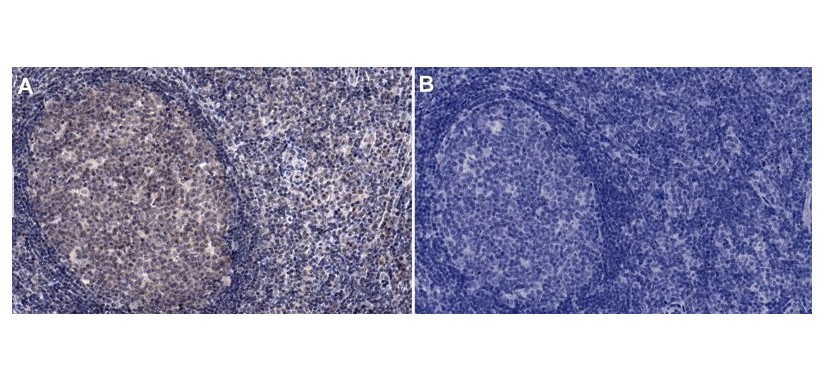
Immunohistochemistry (Paraffin) Analysis: A 1:250 dilution of this antibody
detected Caspase-3 in Human tonsil tissue sections.
Biochem/physiol Actions
Recognizes full-length Caspase 3 (Yama/Apopain) and
proteolytic fragments.
Physical form
Format: Purified
Protein A purified IgG in of 0.1M Tris-glycine, pH 7.4,
0.15M NaCl,and 0.05% sodium azide.
Preparation Note
Stable for 2 years at 2-8°C from date of shipment. For
maximum recovery of product, centrifuge the original vial prior to removing the
cap.
Analysis Note
Control
Positive Antigen Control: Catalog #12-301, non-stimulated A431 cell lysate. Add
2.5µL of 2-mercaptoethanol/100µL of lysate and boil for 5 minutes to reduce the
preparation. Load 20µg of reduced lysate per lane for mingels.
routinely evaluated by immunoblot on RIPA lysates from
non-stimulated human A431 cells, mouse 3T3/A31 or rat PC12 cells
Other Notes
Replaces: 04-1090; 04-439
Legal Information
UPSTATE is a registered trademark of Merck KGaA, Darmstadt,
Germany