General description
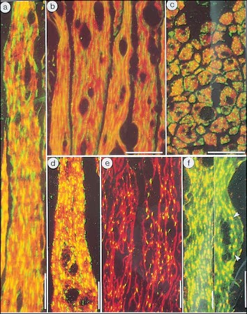
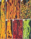
In staining of chicken gizzard ultrathin tissue
cryosections, the antibody labels the dense bodies and longitudinal channels
linking consecutive dense bodies that are also occupied by desmin and the
membrane-associated dense plaque. It does not stain adult cardiac and skeletal
muscles except for traces due to contaminations of the sample with non-muscle
cells, or if embryonic tissue is being used.
Monoclonal Anti-β-Actin (mouse IgG1 isotype) is derived from
the AC-15 hybridoma produced by the fusion of mouse myeloma cells and
splenocytes from an immunized mouse. Actin is one of the most conserved
eukaryotic proteins, it is expressed in mammals and birds as at least six
isoforms. Four of them represent the differentiation markers of muscle tissues
and two are found practically in all cells. There are three α-actins
(α-skeletal, α-cardiac, and α-smooth muscle), one β-actin (β-nonmuscle), and
two γ-actins (γ-smooth muscle and γ-non-muscle). Actin isoforms show >90%
overall sequence homology, but only 50−60% homology in their 18 NH2-terminal
residues. The NH2-terminal region of actin appears to be a major antigenic
region and may be involved in the interaction of actin with other proteins such
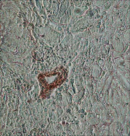
as myosin. The antibody can be used for staining of acetone-fixed frozen
sections, EM preparations, and microinjection experiments. B5, ethanol,
methacam, or Bouin′s solutions can be used as fixatives. The epitope recognized
by the antibody is resistant to formalin-fixed and paraffin-embedding.
Immunogen
slightly modified β-cytoplasmic actin N-terminal peptide,
Ac-Asp-Asp-Asp-Ile-Ala-Ala-Leu-Val-Ile-Asp-Asn-Gly-Ser-Gly-Lys, conjugated to
KLH.
Application
Monoclonal Anti-β-Actin antibody produced in mouse has also
been used:
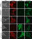
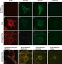
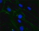
- in
immunofluorescence staining
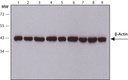
- in
immunoblotting
- in
immunohistochemistry
- as a
control for protein arrays
Monoclonal mouse anti-actin antibody was used as a loading
control for western blot analysis of immunoprecipitated proteins from rat
dorsal root ganglion cocultures.
Monoclonal mouse anti-actin was used as a loading control
for western blot analysis of rat liver protein lysates.
Biochem/physiol Actions
Actin and myosin are constituents of many cell types and are
involved in a myriad of cellular processes including locomotion, cytokinesis,
cytoplasmic streaming, secretion and phagocytosis. The actin in cells of
various species and tissue origin is very similar in its immunological and
physical properties.
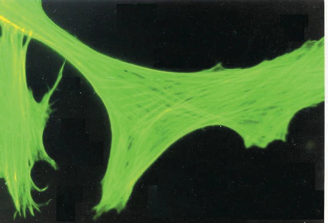
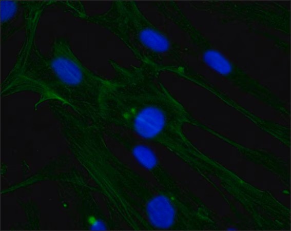
Monoclonal Anti β-Actin antibody recognizes an epitope
located on the N-terminal end of the β-isoform of actin. The antibody
specifically labels β-actin in a wide variety of tissues and species using
immunoblotting (42 kDa), immunofluorescent staining of cultured cell lines, and
immunohistochemistry.
Physical form
Supplied as ascites fluid with 15mM sodium azide as a
preservative.
Other Notes
2024
CiteAb Award Winner for Supplier Succeeding in Parkinson′s Research
To view an Actin antibody selection guide, please
visit www.sigmaaldrich.com/actin.
Disclaimer
Unless otherwise stated in our catalog or other company
documentation accompanying the product(s), our products are intended for
research use only and are not to be used for any other purpose, which includes
but is not limited to, unauthorized commercial uses, in vitro diagnostic uses,
ex vivo or in vivo therapeutic uses or any type of consumption or application
to humans or animals.