General description
Tight junction protein ZO-1 (UniProt Q07157; also known as
Tight junction protein 1, Zona occludens protein 1, Zonula occludens protein 1)
is encoded by the TJP1 gene (also known as ZO1) (Gene ID 7082) in human. The
tight junction (TJ) constitutes the barrier between the apical and the
basolateral domains of the plasma membrane. The assembly and permeability of
this barrier are dependent on the zonula occludens (ZO) membrane-associated
guanylate kinase (MAGUK) proteins ZO-1, ZO-2, and ZO-3. ZO-1, a 210-225 kDa
protein, is found at the submembranous domain of TJs in epithelia and
endothelia. ZO-1 contains three PDZ domains (PDZ1/aa23-109; PDZ2/aa181-261;
PDZ3/aa422-502) at its N-treminal end, followed by an SH3 domain (aa519-580), a
GUK/GK homology domain (aa632-782), an acidic domain (aa817-894), an alpha
spliced domain (aa921-1000), and a C-terminal proline-rich/PR domain. ZO-1 is a
phosphoprotein and a known substrate of serine/threonine kinases ZAK and of
PKC. MAPK signaling pathway regulates tyrosine phosphorylation of ZO-1, and
MEK1 inhibition in Ras transformed epithelial cells is reported to result in
tyrosine phosphorylation of ZO-1 and occludin. ZO-1 interacts with claudins,
JAM, ZO-2, and ZO-3 through its PDZ domains, while its GK module mediates interaction
with occludin. ZO-1 also binds actin cytoskeleton and actin-binding protein 4.1
through its carboxyl terminal end. In addition, ZO-1 associates with AF-6 and
cingulin at the TJ, as well as with the adherens junction protein alpha-catenin
and with the gap junction proteins connexins 43 and 45.
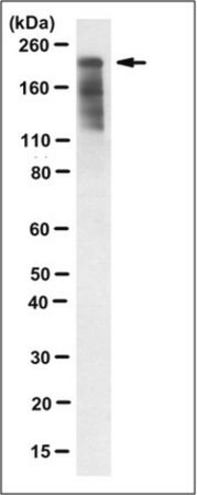
~195 kDa observed
Immunogen
GST-tagged recombinant protein corresponding to human ZO-1.
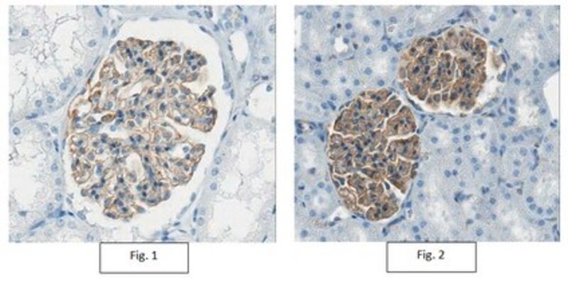
Application
Immunohistochemistry Analysis: A 1:1,000 dilution from a
representative lot detected ZO-1 in human and rat kidney tissues.
Research Category
Cell Structure
Research Sub Category
Adhesion (CAMs)
This Anti-ZO-1 Antibody, clone 5G6.1 is validated for use in
Western Blotting, Immunocytochemistry, Flow Cytometry, ELISA for the detection
of ZO-1 .
Physical form
Format: Purified
Protein G Purified
Purified mouse monoclonal IgG2aκ antibody in buffer
containing 0.1 M Tris-Glycine (pH 7.4), 150 mM NaCl with 0.05% sodium azide.
Preparation Note
Stable for 1 year at 2-8°C from date of receipt.
Analysis Note
Evaluated by Western Blotting in HCT116 cell lysate.
Western Blotting Analysis: 0.5 µg/mL of this antibody detected ZO-1 in 200 µg
of HCT116 cell lysate.
Other Notes
Concentration: Please refer to lot specific datasheet.
Disclaimer
Unless otherwise stated in our catalog or other company
documentation accompanying the product(s), our products are intended for
research use only and are not to be used for any other purpose, which includes
but is not limited to, unauthorized commercial uses, in vitro diagnostic uses,
ex vivo or in vivo therapeutic uses or any type of consumption or application
to humans or animals.