General description
Actin, cytoplasmic 1 (UniProt P60709; also known as
Beta-actin) is encoded by the ACTB gene (Gene ID 60) in human. Actins are
globular multi-functional proteins that serve as the basic building blocks of
cytoskeletal microfilaments and are among the most conserved eukaryotic
proteins. Six actin types exist, skeletal muscle alpha-actin is encoded by the
ACTA1 gene, smooth muscle alpha-actin by the ACTA2 gene, cytoplasmic beta-actin
by the ACTB gene, cardiac muscle alpha-actin by the ACTC1 gene, cytoplasmic gamma-actin
by the ACTG1 gene, and smooth muscle gamma-actin by the ACTG2 (a.k.a. ACTA3)
gene. Although actins show >90% overall sequence homology, isoforms do show
spatial, temporal, and tissue-specific expression patterns and only 50-60%
homology is found in their 18 N-terminal residues. Cytoplasmic β and γ-actins,
are thought to be present in all cells, while the other four actin isoforms are
typically found in specific adult muscle tissue types. Actins exist in a
variety of structural states, depending on the specific ionic conditions or the
interaction with ligand proteins. The oligomeric and polymeric forms that actin
molecules assume are dependent on the distinct conformations they adopt.
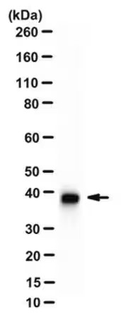
~39-45 kDa observed. Uncharacterized band(s) may appear in
some lysates.
Immunogen
KLH-conjugated linear peptide corresponding to the
N-terminal sequence of human beta-Actin.
Application
This Anti-beta-Actin Antibody, clone 4C2 is validated for
use in Western Blotting, Immunocytochemistry for the detection of beta-Actin.
Western Blotting Analysis: 0.5 µg/mL from a representative
lot detected beta-Actin in 10 µg of NIH/3T3 cell lysate.
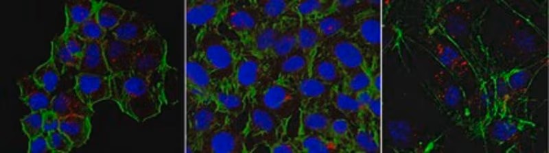
Immunocytochemistry Analysis: 5.0 µg/mL from a representative lot detected
beta-Actin in HUVECs, A431 and HeLa cells.
Western Blotting Analysis: A representative lot detected downregulated
beta-actin levels in stimulated (by A23187, TRAP-6, TNF, LPS, or IFN-γ) human
cerebral microvascular endothelial D3 cells (hCMEC/D3) and their microparticles
(MPs) when compared with unstimulated hCMEC/D3 and their MPs (Latham, S.L., et
al. (2013). FASEB J. 27(2):672-683).
Western Blotting Analysis: A representative lot detected siRNA-mediated
downregulation of beta-actin in A549 human lung carcinoma cells (Miazza, V., et
al. (2011). Virology. 410(1):7-16).
Western Blotting Analysis: A representative lot detected beta-actin, but not
cytoplasmic gamma-actin separated by 2-D gel electrophoresis of purified
chicken gizzard actins or total protein extracts from human subcutaneous
fibroblasts (HSCFs), canine MDCK cells, and rat aorta tissue (Dugina, V., et
al. (2009). J. Cell Sci. 122(Pt 16):2980-2988).
Western Blotting Analysis: A representative lot detected BSA conjugated with
beta-actin N-terminal peptide, but not BSA conjugated with N-terminal peptides
derived from the other 5 actin types (Dugina, V., et al. (2009). J. Cell Sci.
122(Pt 16):2980-2988).
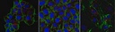
Immunocytochemistry Analysis: A representative lot detected TNF-stimulated
localization of β-actin into thick, intensely staining stress fibers prominent
at the basal surface of of human cerebral microvascular endothelial D3 cells
(hCMEC/D3). Rho kinase inhibitor Y-27632 (Cat. No. 688000) treatment suppressed
TNF-induced β-actin stress fiber formation (Latham, S.L., et al. (2013). FASEB
J. 27(2):672-683).
Immunocytochemistry Analysis: A representative lot detected a drastic
subcellular redistribution of beta-actin following Sendai virus infection of
polarized Madin-Darby canine kidney (MDCK) epithelial cells by fluorescent
immunocytochemistry staining of paraformaldehyde-fixed, methanol-treated cells
(Miazza, V., et al. (2011). Virology. 410(1):7-16).
Immunocytochemistry Analysis: A representative lot detected beta-actin
subcellular localization distinct from that of cytoplasmic gamma-actin in both
spreading and stationary cells by fluorescent immunocytochemistry, using
paraformaldehyde-fixed, methanol-treated HSCF human subcutaneous fibroblasts,
HaCaT human keratinocytes, WI38 human embryonic fibroblasts,and Madin-Darby
canine kidney (MDCK) cells (Dugina, V., et al. (2009). J. Cell Sci. 122(Pt
16):2980-2988).
Biochem/physiol Actions
Clone 4C2 detected BSA conjugated with beta-actin N-terminal
peptide, but not BSA conjugated with N-terminal peptides derived from the 5
other actin types (Dugina, V., et al. (2009). J. Cell Sci. 122(Pt
16):2980-2988).
Physical form
Format: Purified
Analysis Note
Evaluated by Western Blotting in HeLa cell lysate.
Western Blotting Analysis: 0.5 µg/mL of this antibody detected beta-Actin in 10
µg of HeLa cell lysate.
Other Notes
Concentration: Please refer to lot specific datasheet.